
What is Nuclear Medicine?
Nuclear Medicine is a medical specialty that utilizes safe, non-invasive, and cost-efficient methods to both visualize internal body structures and manage various diseases.
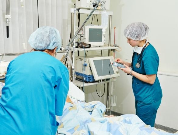
Nuclear imaging is unique and documents both organ function and morphology. Gamma ray emitting radiotracer is injected intravenously and non-invasive imaging is performed with state-of-the-art Dual Head Gamma Camera (GE Millennium VG)
Nuclear Medicine is a medical speciality that uses safe, painless, and cost-effective
techniques both to image the body and treat disease.
Nuclear imaging is unique in that it documents both organ function and morphology.Gamma ray emitting radiotracer is injected intravenously and non-invasive imaging is performed with state-of-the-art Dual Head Gamma Camera (GE Millennium VG)
When we already have X-ray, Ultrasound, CT & MRI, why do we need this?
- It provides unique functional information from cellular and molecular levels which is not possible with other modalities.
- It is an extremely sensitive technique
- It provides dynamic and quantitative information
- It enables objective and prognostic assessment of disease
Radionuclide procedures are available for almost all organ systems just like the different X-ray procedures. It is used in evaluation of Heart, Lung, Brain, Bone, Kidneys, Liver etc.Scans are designed to study various aspects like concentration, excretion, drainage/flow of tracers in various organs or localize/characterize the leison pathology.
- Nuclear Scan is very safe
- There will be no reaction or side effect to the tracer injected
- Radiation burden to the patient’s body is less than in X-ray Procedures
PET CT

GE/Discovery IQ PET CT – First in South Tamilnadu
PET CT is revolutionizing Cancer care
The value of Whole Body Positron Emission Tomography–Computed Tomography (PET/CT) for diagnostic imaging in oncology is well established, with its capabilities to combine anatomical and functional aspects of the whole body to provide a complete picture of the patient’s disease status.
Most common uses of PET/CT are found in: Oncology, Cardiology, and Neurology practices.
FDG PET/CT in Oncology
FDG PET/CT is an integral part of cancer management in evidence-based practice. It is used for initial staging of various cancers, radiotherapy planning, treatment response assessment, restaging, and surveillance. It changes treatment decisions in a significant proportion of cancer patients.
FDG PET/CT in Cardiology
In cardiology practice, FDG PET/CT is the gold standard test for detection of myocardial viability. It provides crucial evidence for selecting coronary artery disease patients for revascularization (stenting or bypass surgery).
FDG PET/CT in Neurology
In neurology practice, FDG PET/CT is used to provide differential diagnosis for dementia. It helps neurosurgeons in brain mapping for surgical resection in cases of drug-resistant epilepsy.
Stress Myocardial Perfusion SPECT
GE/Discovery IQ PET CT – First in South Tamilnadu
PET CT is revolutionizing Cancer care
The value of Whole Body Positron Emission Tomography–Computed Tomography (PET/CT) for diagnostic imaging in oncology is well established, with its capabilities to combine anatomical and functional aspects of the whole body to provide a complete picture of the patient’s disease status.
Most common uses of PET/CT are found in: Oncology, Cardiology, and Neurology practices.
FDG PET/CT in Oncology
FDG PET/CT is an integral part of cancer management in evidence-based practice. It is used for initial staging of various cancers, radiotherapy planning, treatment response assessment, restaging, and surveillance. It changes treatment decisions in a significant proportion of cancer patients.
FDG PET/CT in Cardiology
In cardiology practice, FDG PET/CT is the gold standard test for detection of myocardial viability. It provides crucial evidence for selecting coronary artery disease patients for revascularization (stenting or bypass surgery).
FDG PET/CT in Neurology
In neurology practice, FDG PET/CT is used to provide differential diagnosis for dementia. It helps neurosurgeons in brain mapping for surgical resection in cases of drug-resistant epilepsy.
Clinical Indications
DTPA Renal Scan
- Split renal functions
- GFR estimation
- Evaluation of hydronephrosis
- PUJ stenosis
- Renovascular Hypertension
- Transplant evaluation
DMSA Cortical Scan
- Pyelonephritis
- Urinary tract infection
- Reflux disease
- Ectopic Kidney
Voiding Cystography
- Detection & follow-up of ureteric reflux
Gastro Enterology
Hepatobilliary Scan
- To diagnose acute cholecystitis
- To assess Gall Bladder dysfunction
- To study bile drainage, atresia and post OP cases
Liver & Spleen Scan
- To evaluate cirrhosis
- Buddchiari, Nodular Hyperplasia and tumors
Other Scans
- Hemangioma of the Liver
- GI bleeding
- Ectopic Gastric Mucosa
Meckel’s diverticulum
- Oesophageal transit in dysphagia
- GE Reflux scintigraphy
- Gastic Emptying in Gastroparesis, post OP states, dysmotility etc.
Gamma Camera – SPECT
Thallium Scan
Myocardial Perfusion Scan visualizes the distribution of tracer uptake in the heart muscle, which reflects regional blood flow in different coronary artery territories. This can be performed with 201T1 / 99m Tc-Sestamibi / Trofosmin.
Rest Injection Scan indicates whether the heart muscle is viable or scarred due to prior attacks.
Stress (Exercise/Pharmacological) Scan reveals inducible ischemic perfusion defects corresponding to significant coronary disease.
Brain Perfusion SPECT with advanced NeuroGam Analysis Package for diagnosing
- Assessment of stroke
- To detect Epileptic focus
- To evaluate dementia
Other Indications
- Thyroid Scan to evaluate palpable thyroid nodule, midline neck swelling and thyromegaly & toxic goitres
- Radio Iodine Scan for post operative thyroid cancer
- Lung Ventilation Perfusion Scan for pulmonary embolism
- Radionuclide Ventriculography for LV function & Ejection fraction
- Isotope Venography for DVT
- Scinti-mammography for doubtful breast leison
Advanced Cardiac Analysis with Quantitative Perfusion
SPECT and Gated SPECT
SPECT and Gated SPECT software provides the following information:
- Universal slice review
- Bullseye plot
- Cardiac ejection fraction
- Wall motion abnormality
- Myocardial viability
Clinical Indications
- Detection of Coronary Artery Disease
- Chest pain evaluation – Stress test with uninterpretable ECG
- Assessment of borderline coronary stenosis seen in Angiography
- Assessment of myocardial viability
- Cardiac fitness for non-cardiac surgery
Whole Body Bone Scan
A bone scan is a diagnostic imaging study which records the distribution of a radioactive tracer in the skeletal system. It is the most sensitive study available to pick up any pathology of the skeleton.
Clinical Indications
- Metastatic bone lesions
- Primary bone lesions
- Osteoid osteoma
- Infection/Inflammation
- Osteomyelitis
- Sacroilitis
- Avascular necrosis
- Trauma stress fracture
- Metabolic bone disease
- Bone pain evaluation – low back ache evaluation
Renal Scan
Renal scans are performed to evaluate the differential renal function, the extraction and excretion function of the kidneys. Information about Glomerular Filtration Rate (GFR), tubular function, and cortical morphology can be obtained by such scans.
Brain SPECT
Brain Perfusion SPECT with advanced NeuroGam Analysis Package for diagnosing
- Assessment of stroke
- To detect Epileptic focus
- To evaluate dementia
Other Indications
- Thyroid Scan to evaluate palpable thyroid nodule, midline neck swelling and thyromegaly & toxic goitres
- Radio Iodine Scan for post operative thyroid cancer
- Lung Ventilation Perfusion Scan for pulmonary embolism
- Radionuclide Ventriculography for LV function & Ejection fraction
- Isotope Venography for DVT
- Scinti-mammography for doubtful breast leison
Our Doctors (Madurai)
Our Doctors (Dindigul)

If you have questions, feel free to reach out
Nuclear Medicine uses small amounts of radioactive materials to visualize organ function and detect diseases at a molecular level, unlike regular imaging that mainly shows structural details. This helps in early diagnosis and personalized treatment.
Yes, Nuclear Medicine is safe. The radioactive tracers used are minimal, carefully controlled, and typically eliminated from the body quickly. Our team ensures patient safety throughout the procedure.
Molecular Imaging helps diagnose cancer, heart diseases, neurological disorders, and infections by showing cellular activity, enabling precise treatment planning and monitoring.